|
Nittobo America
rabbit polyclonal antibody against cnr1 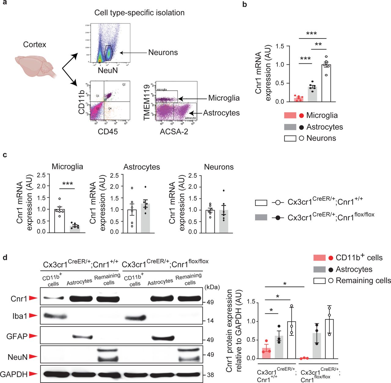 Rabbit Polyclonal Antibody Against Cnr1, supplied by Nittobo America, used in various techniques. Bioz Stars score: 90/100, based on 1 PubMed citations. ZERO BIAS - scores, article reviews, protocol conditions and more https://www.bioz.com/result/rabbit polyclonal antibody against cnr1/product/Nittobo America Average 90 stars, based on 1 article reviews
rabbit polyclonal antibody against cnr1 - by Bioz Stars,
2026-03
90/100 stars
|
Buy from Supplier |
|
Interchim Chemicals
syp rabbit polyclonal rb-1461-p1 antibody  Syp Rabbit Polyclonal Rb 1461 P1 Antibody, supplied by Interchim Chemicals, used in various techniques. Bioz Stars score: 90/100, based on 1 PubMed citations. ZERO BIAS - scores, article reviews, protocol conditions and more https://www.bioz.com/result/syp rabbit polyclonal rb-1461-p1 antibody/product/Interchim Chemicals Average 90 stars, based on 1 article reviews
syp rabbit polyclonal rb-1461-p1 antibody - by Bioz Stars,
2026-03
90/100 stars
|
Buy from Supplier |
|
bioMerieux gmbh
anti-ns4 rabbit polyclonal antibody rb Anti Ns4 Rabbit Polyclonal Antibody Rb, supplied by bioMerieux gmbh, used in various techniques. Bioz Stars score: 90/100, based on 1 PubMed citations. ZERO BIAS - scores, article reviews, protocol conditions and more https://www.bioz.com/result/anti-ns4 rabbit polyclonal antibody rb/product/bioMerieux gmbh Average 90 stars, based on 1 article reviews
anti-ns4 rabbit polyclonal antibody rb - by Bioz Stars,
2026-03
90/100 stars
|
Buy from Supplier |
|
Spring Bioscience
anti-human polyclonal rabbit antibodies rb anti-bak pab Anti Human Polyclonal Rabbit Antibodies Rb Anti Bak Pab, supplied by Spring Bioscience, used in various techniques. Bioz Stars score: 90/100, based on 1 PubMed citations. ZERO BIAS - scores, article reviews, protocol conditions and more https://www.bioz.com/result/anti-human polyclonal rabbit antibodies rb anti-bak pab/product/Spring Bioscience Average 90 stars, based on 1 article reviews
anti-human polyclonal rabbit antibodies rb anti-bak pab - by Bioz Stars,
2026-03
90/100 stars
|
Buy from Supplier |
|
Interchim Chemicals
rabbit polyclonal antibody against human smooth muscle -actin rb-9010-p Rabbit Polyclonal Antibody Against Human Smooth Muscle Actin Rb 9010 P, supplied by Interchim Chemicals, used in various techniques. Bioz Stars score: 90/100, based on 1 PubMed citations. ZERO BIAS - scores, article reviews, protocol conditions and more https://www.bioz.com/result/rabbit polyclonal antibody against human smooth muscle -actin rb-9010-p/product/Interchim Chemicals Average 90 stars, based on 1 article reviews
rabbit polyclonal antibody against human smooth muscle -actin rb-9010-p - by Bioz Stars,
2026-03
90/100 stars
|
Buy from Supplier |
|
MyBiosource Biotechnology
rabbit polyclonal to pv msp1 antibody rb-pabv  Rabbit Polyclonal To Pv Msp1 Antibody Rb Pabv, supplied by MyBiosource Biotechnology, used in various techniques. Bioz Stars score: 90/100, based on 1 PubMed citations. ZERO BIAS - scores, article reviews, protocol conditions and more https://www.bioz.com/result/rabbit polyclonal to pv msp1 antibody rb-pabv/product/MyBiosource Biotechnology Average 90 stars, based on 1 article reviews
rabbit polyclonal to pv msp1 antibody rb-pabv - by Bioz Stars,
2026-03
90/100 stars
|
Buy from Supplier |
|
Becton Dickinson
anti-rb rabbit polyclonal antibody 554136 Anti Rb Rabbit Polyclonal Antibody 554136, supplied by Becton Dickinson, used in various techniques. Bioz Stars score: 90/100, based on 1 PubMed citations. ZERO BIAS - scores, article reviews, protocol conditions and more https://www.bioz.com/result/anti-rb rabbit polyclonal antibody 554136/product/Becton Dickinson Average 90 stars, based on 1 article reviews
anti-rb rabbit polyclonal antibody 554136 - by Bioz Stars,
2026-03
90/100 stars
|
Buy from Supplier |
|
WuXi AppTec
polyclonal rabbit anti-human antibody slug (rb 1398 Polyclonal Rabbit Anti Human Antibody Slug (Rb 1398, supplied by WuXi AppTec, used in various techniques. Bioz Stars score: 90/100, based on 1 PubMed citations. ZERO BIAS - scores, article reviews, protocol conditions and more https://www.bioz.com/result/polyclonal rabbit anti-human antibody slug (rb 1398/product/WuXi AppTec Average 90 stars, based on 1 article reviews
polyclonal rabbit anti-human antibody slug (rb 1398 - by Bioz Stars,
2026-03
90/100 stars
|
Buy from Supplier |
|
Becton Dickinson
bax (rb pab, rabbit polyclonal antibody  Bax (Rb Pab, Rabbit Polyclonal Antibody, supplied by Becton Dickinson, used in various techniques. Bioz Stars score: 90/100, based on 1 PubMed citations. ZERO BIAS - scores, article reviews, protocol conditions and more https://www.bioz.com/result/bax (rb pab, rabbit polyclonal antibody/product/Becton Dickinson Average 90 stars, based on 1 article reviews
bax (rb pab, rabbit polyclonal antibody - by Bioz Stars,
2026-03
90/100 stars
|
Buy from Supplier |
|
Cambridge Bioscience
antibody cf633 anti-rb (donkey anti-rabbit polyclonal)  Antibody Cf633 Anti Rb (Donkey Anti Rabbit Polyclonal), supplied by Cambridge Bioscience, used in various techniques. Bioz Stars score: 90/100, based on 1 PubMed citations. ZERO BIAS - scores, article reviews, protocol conditions and more https://www.bioz.com/result/antibody cf633 anti-rb (donkey anti-rabbit polyclonal)/product/Cambridge Bioscience Average 90 stars, based on 1 article reviews
antibody cf633 anti-rb (donkey anti-rabbit polyclonal) - by Bioz Stars,
2026-03
90/100 stars
|
Buy from Supplier |
|
Medicorp Inc Canada
anti-nm23 rabbit polyclonal antibody rb-116 Anti Nm23 Rabbit Polyclonal Antibody Rb 116, supplied by Medicorp Inc Canada, used in various techniques. Bioz Stars score: 90/100, based on 1 PubMed citations. ZERO BIAS - scores, article reviews, protocol conditions and more https://www.bioz.com/result/anti-nm23 rabbit polyclonal antibody rb-116/product/Medicorp Inc Canada Average 90 stars, based on 1 article reviews
anti-nm23 rabbit polyclonal antibody rb-116 - by Bioz Stars,
2026-03
90/100 stars
|
Buy from Supplier |
|
Full Moon BioSystems
polyclonal rabbit antibody to phospho-rb ser795 Polyclonal Rabbit Antibody To Phospho Rb Ser795, supplied by Full Moon BioSystems, used in various techniques. Bioz Stars score: 90/100, based on 1 PubMed citations. ZERO BIAS - scores, article reviews, protocol conditions and more https://www.bioz.com/result/polyclonal rabbit antibody to phospho-rb ser795/product/Full Moon BioSystems Average 90 stars, based on 1 article reviews
polyclonal rabbit antibody to phospho-rb ser795 - by Bioz Stars,
2026-03
90/100 stars
|
Buy from Supplier |
Image Search Results
Journal: bioRxiv
Article Title: Microglial cannabinoid receptor type 1 mediates social memory deficits produced by adolescent THC exposure and 16p11.2 duplication
doi: 10.1101/2023.07.24.550212
Figure Lengend Snippet: a, Experimental flow of Fluorescence-Activated Cell Sorting (FACS)-based microglia (CD45 + CD11b + TMEM119 + ), astrocyte (ACSA-2 + ), and neuron (NeuN + ) isolation using specific cell markers. b, Relative mRNA expression levels of Cnr1 in microglia, astrocytes, and neurons isolated from wild type mice were measured by quantitative real time PCR (qPCR) using the TaqMan assay protocol. n = 6 mice per group. *** p < 0.001, ** p < 0.01, determined by one-way ANOVA with post hoc Tukey test. c, Relative mRNA expression levels of Cnr1 in microglia, astrocytes, and neurons isolated from Cx3cr1 CreER/+ ; Cnr1 +/+ and Cx3cr1 CreER/+ ; Cnr1 flox/flox mice were measured by qPCR. n = 6 mice per group. *** p < 0.001, determined by Student t -test. d, Microglia-enriched CD11b + cells, ACSA-2 + astrocytes, and remaining cells including neurons were collected from the cerebral cortex of Cx3cr1 CreER/+ ; Cnr1 flox/flox mice and littermate controls ( Cx3cr1 CreER/+ ; Cnr1 +/+ ) and by magnetic activated cell sorting (MACS). For each cell type, expression of Cnr1, marker proteins (Iba1, GFAP, NeuN) and a loading control (GAPDH) in the total protein were analyzed with SDS-PAGE followed by Western blotting with 10 μg of protein sample loaded in each well. n = 3 mice per group. * p < 0.05, determined by two-way ANOVA with post hoc Tukey test. (b, c, d) Data are presented as the mean ± s.e.m.
Article Snippet: The following primary antibodies were used:
Techniques: Fluorescence, FACS, Isolation, Expressing, Real-time Polymerase Chain Reaction, TaqMan Assay, Marker, SDS Page, Western Blot

Journal: bioRxiv
Article Title: Microglial cannabinoid receptor type 1 mediates social memory deficits produced by adolescent THC exposure and 16p11.2 duplication
doi: 10.1101/2023.07.24.550212
Figure Lengend Snippet: a , Apoptosis assay of primary microglia cultures produced from WT and 16p11dup male mice. Quantification of signal intensity of apoptosis marker apopxin ( n = 12 fields in 3 mice per condition). b, Necrosis assay of primary microglia cultures produced from WT and 16p11dup male mice. Quantification of signal intensity of necrosis marker 7-AAD ( n = 12 fields in 3 mice per condition). c, Quantification of cellular process area of phalloidin-stained microglia cultures produced from WT and 16p11dup male mice ( n = 12 fields in 3 mice per condition). d , Quantification of cellular process number of phalloidin-stained microglia cultures produced from WT and 16p11dup male mice ( n = 12 fields in 3 mice per condition). e, Representative images of microglia cell cultures produced from WT and 16p11dup male mice in apoptosis and necrosis assays. Apopxin (green) and 7-AAD (red) are shown. Scale bar, 50 μm. f, Immunohistochemistry with antibody against phalloidin (green) of primary microglia cultures produced from WT and 16p11dup male mice. Scale bar, 25 μm. g , Apoptosis assay of primary microglia cultures produced from genetic deletion of Cnr1 (Cnr1 KO) and genetic deletion of Cnr2 (Cnr2 KO) male mice. Quantification of signal intensity of apoptosis marker apopxin ( n = 12 fields in 3 mice per condition). h, Necrosis assay of primary microglia cultures produced from Cnr1 KO and Cnr2 KO male mice. Quantification of signal intensity of necrosis marker 7-AAD ( n = 12 fields in 3 mice per condition). i, Quantification of cellular process area of phalloidin-stained microglia cultures produced from Cnr1 KO and Cnr2 KO male mice. ( n = 12 fields in 3 mice per condition). j , Quantification of cellular process number of phalloidin-stained microglia cultures of Cnr1 KO and Cnr2 KO male mice ( n = 12 fields in 3 mice per condition). k, Representative images of microglia cell cultures produced from Cnr1 KO and Cnr2 KO male mice in apoptosis and necrosis assays. Apopxin (green) and 7-AAD (red) are shown. Scale bar, 100 μm. l, Immunohistochemistry with antibody against phalloidin (green) of primary microglia cultures of Cnr1 KO and Cnr2 KO male mice. Scale bar, 25 μm. m, Representative images of immunohistochemistry of Iba1 (green) and Casp3-p17 (red) (top left) as well as TUNEL signals (red) and DAPI (blue) (bottom left) in the mPFC at P51. Scale bar, 50 μm. Quantification of signal intensity of Casp3-p17 and TUNEL (right) ( n = 6 mice per group). ( a, c, d, m ) *** p < 0.001, ** p < 0.01, * p < 0.05, determined by two-way ANOVA with post hoc Tukey test. ( g, i, j ) *** p < 0.001, ** p < 0.01, determined by Student t -test. Data are presented as the mean ± s.e.m.
Article Snippet: The following primary antibodies were used:
Techniques: Apoptosis Assay, Produced, Marker, Staining, Immunohistochemistry, TUNEL Assay

Journal: bioRxiv
Article Title: Microglial cannabinoid receptor type 1 mediates social memory deficits produced by adolescent THC exposure and 16p11.2 duplication
doi: 10.1101/2023.07.24.550212
Figure Lengend Snippet: a, Schematic diagram of the experimental design. 16p11 dup ; Cx3cr1 CreER/+ ; Cnr1 +/+ , 16p11 dup ; Cx3cr1 CreER/+ ; Cnr1 flox/flox , wild type littermate (16p11 wt ); Cx3cr1 CreER/+ ; Cnr1 +/+ , and 16p11 wt ; Cx3cr1 CreER/+ ; Cnr1 flox/flox mice were given tamoxifen (0.1 mg/g body weight) orally once a day for 5 consecutive days at P21-P25, 5 days before the start of THC (s.c., 8mg/kg) treatment during adolescence (P30-P51), followed by microglial phenotyping at P51 (upon completion of THC treatment). b, Immunohistochemical analysis of Iba1 (green) in the mPFC at P51. (Left) Representative images of the mPFC, representative tracing images (red), and images of cellular processes (green) and cell bodies (yellow) of Iba1 + cells. Scale bar, 50 μm (left) and 10 μm (middle and right). c, The number of Iba1 + P2ry12 + cells (left) and Iba1 + P2ry12 - cells (middle) in the mPFC, presented as % of control. (Right) The percentage of Iba1 + P2ry12 - cells among all Iba1 + cells in the mPFC. ( n = 6 slices in 3 mice per condition). d, Quantification of the ratio of cellular process area (left) and cell body size to total cell size (right) of Iba1 + cells. ( n = 30 cells in 3 mice per condition). e, Representative images of immunohistochemistry of Iba1 (green) and Casp3-p17 (red) (top left) as well as TUNEL signals (red) and DAPI (blue) (bottom left) in the mPFC at P51. Scale bar, 50 μm. f , Quantification of signal intensity of Casp3-p17 in Iba1 + cells (left) and TUNEL (right). n = 6 slices in 3 mice per condition. ( c, d, f ) *** p < 0.001, ** p < 0.01, * p < 0.05, determined by two-way ANOVA with post hoc Tukey test. Data are presented as the mean ± s.e.m.
Article Snippet: The following primary antibodies were used:
Techniques: Immunohistochemical staining, Immunohistochemistry, TUNEL Assay

Journal: bioRxiv
Article Title: Microglial cannabinoid receptor type 1 mediates social memory deficits produced by adolescent THC exposure and 16p11.2 duplication
doi: 10.1101/2023.07.24.550212
Figure Lengend Snippet: Cnr1 deletion in the microglia normalizes deficits in PT neurons and social memory that are synergistically produced by adolescent THC treatment and 16p11dup. a , Schematic diagram of the experimental design. 16p11 dup ; Cx3cr1 CreER/+ ; Cnr1 +/+ , 16p11 dup ; Cx3cr1 CreER/+ ; Cnr1 flox/ flox , wild type littermate (16p11 wt ); Cx3cr1 CreER/+ ; Cnr1 +/+ , and 16p11 wt ; Cx3cr1 CreER/+ ; Cnr1 flox/flox mice were given tamoxifen (0.1 mg/g body weight) orally once a day for 5 consecutive days at P21-P25, 5 days before the start of THC (s.c., 8mg/kg) treatment during adolescence (P30-P51), followed by electrophysiological experiments or behavioral assays at P72 (after a 3-week abstinence period at adulthood). b, (Left) Representative voltage traces recorded from PT neurons in response to current step injections in the presence of blockers for AMPA, NMDA, and GABAa receptors. (Right) The intrinsic excitability of PT neurons, as quantified by input resistance (left), rheobase (middle), and spike frequency (right). 16p11 wt ; Cx3cr1 CreER/+ ; Cnr1 +/+ ( n = 9 cells in 2 mice), 16p11 wt ; Cx3cr1 CreER/+ ; Cnr1 flox/flox ( n = 5 cells in 3 mice), 16p11 dup ; Cx3cr1 CreER/+ ; Cnr1 +/+ ( n = 6 cells in 2 mice), and 16p11 dup ; Cx3cr1 CreER/+ ; Cnr1 flox/flox ( n = 8 cells in 3 mice). ** p < 0.01, * p < 0.05, determined by two-way ANOVA with post hoc Tukey test. c, (Left) Representative voltage traces recorded from IT neurons in response to current step injections in the presence of blockers for AMPA, NMDA, and GABAa receptors. (Right) The intrinsic excitability of IT neurons, as quantified by input resistance (left), rheobase (middle), and spike frequency (right). 16p11 wt ; Cx3cr1 CreER/+ ; Cnr1 +/+ ( n = 4 cells in 2 mice), 16p11 wt ; Cx3cr1 CreER/+ ; Cnr1 flox/flox ( n = 4 cells in 2 mice), 16p11 dup ; Cx3cr1 CreER/+ ; Cnr1 +/+ ( n = 6 cells in 2 mice), and 16p11 dup ; Cx3cr1 CreER/+ ; Cnr1 flox/flox ( n = 5 cells in 2 mice). d, Sociability phenotypes (left) and preference of social novelty (right) as indicated by the discrimination index in the three-chamber social interaction test. e, Time spent with an ovariectomized female mouse in the 5-trial social memory test. ( d , e ) 16p11 wt ; Cx3cr1 CreER/+ ; Cnr1 +/+ ( n = 6 mice), 16p11 wt ; Cx3cr1 CreER/+ ; Cnr1 flox/flox ( n = 5 mice), 16p11 dup ; Cx3cr1 CreER/+ ; Cnr1 +/+ ( n = 6 mice), 16p11 dup ; Cx3cr1 CreER/+ ; Cnr1 flox/flox ( n = 6 mice). ( d, e ) *** p < 0.001, ** p < 0.01, determined by two-way ANOVA with post hoc Tukey test. Data are presented as the mean ± s.e.m.
Article Snippet: The following primary antibodies were used:
Techniques: Produced
Journal: International Journal of Molecular Sciences
Article Title: Cerebral Benefits Induced by Electrical Muscle Stimulation: Evidence from a Human and Rat Study
doi: 10.3390/ijms25031883
Figure Lengend Snippet: Primary antibodies.
Article Snippet:
Techniques: Recombinant, Transduction, Purification
Journal: Sensors (Basel, Switzerland)
Article Title: A Novel Carbon Nanofibers Grown on Glass Microballoons Immunosensor: A Tool for Early Diagnosis of Malaria
doi: 10.3390/s140814686
Figure Lengend Snippet: Glass slides showing ( a ) visual signal obtained at the first capture zone from the detection of Pf HRP-2 in PBS; ( b ) signal observed at the second capture zone from the detection of Pv MSP-1 in solution; and ( c ) signals obtained at the Pf HRP-2 and Pv MSP-1 capture zones from a mixed Pf HRP-2 and Pv MSP-1 solution.
Article Snippet: The following chemicals were used for the study: polyethylene glycol solution (PEG, M W = 8000), glycerol, and bovine serum albumin (BSA) were purchased from Sigma-Aldrich (St. Louis, MO, USA); phosphate buffered saline 1× solution (PBS), 1-ethyl-3-(3-dimethylaminopropyl) carbodiimide hydrochloride (EDC), and N -hydroxysulfosuccinimide (sulfo-NHS) obtained from Thermo Fisher Scientific (Waltham, MA, USA); Plasmodium falciparum histidine rich protein-2 ( Pf HRP-2) antigen, Pv MSP-1, mouse monoclonal to
Techniques:

Journal: Sensors (Basel, Switzerland)
Article Title: A Novel Carbon Nanofibers Grown on Glass Microballoons Immunosensor: A Tool for Early Diagnosis of Malaria
doi: 10.3390/s140814686
Figure Lengend Snippet: ( a ) Variation of percent area of the capture zone covered with NMBs against Pf MSP-1 concentrations and ( b ) Variation of the resistivity of the capture zone with the concentration of immobilized Pf MSP-1. The error bars in both graphs are also shown.
Article Snippet: The following chemicals were used for the study: polyethylene glycol solution (PEG, M W = 8000), glycerol, and bovine serum albumin (BSA) were purchased from Sigma-Aldrich (St. Louis, MO, USA); phosphate buffered saline 1× solution (PBS), 1-ethyl-3-(3-dimethylaminopropyl) carbodiimide hydrochloride (EDC), and N -hydroxysulfosuccinimide (sulfo-NHS) obtained from Thermo Fisher Scientific (Waltham, MA, USA); Plasmodium falciparum histidine rich protein-2 ( Pf HRP-2) antigen, Pv MSP-1, mouse monoclonal to
Techniques: Concentration Assay
Journal: Cell Cycle
Article Title: Curcumin induces Apaf-1-dependent, p21-mediated caspase activation and apoptosis
doi: 10.4161/cc.10.23.18292
Figure Lengend Snippet: Curcumin induces Bax-dependent, p21-mediated cytochrome c release without p53 requirement. HCT116 WT (A), HCT116-p53-/- (B), HCT116-Bax-/- (C) and HCT116-p21-/- (D) cells were treated with curcumin (15 µM) for the indicated times. Cytosolic and mitochondrial fractions were isolated and equal amounts of protein were subjected to protein gel blotting for the detection of cytochrome c (Cyt. C), cytochrome c oxidase subunit II (COX II), heat shock protein 60 (Hsp60) and actin. Actin and Hsp60 serve as loading controls. *represents a non-specific band.
Article Snippet: Primary antibodies were anti-cytochrome c (mAb, monoclonal antibody), -Apaf-1 and -
Techniques: Isolation
Journal: eLife
Article Title: Unrestrained growth of correctly oriented microtubules instructs axonal microtubule orientation
doi: 10.7554/eLife.77608
Figure Lengend Snippet:
Article Snippet: Antibody ,
Techniques: Mutagenesis, Recombinant, Software